Foot Muscles Mri Anatomy - Mri Of The Ankle Detailed Anatomy W Radiology - First of all they act upon the metatarsophalangeal joint of the big toe, leading to the abduction (abductor hallucis muscle), adduction (adductor hallucis muscle) and flexion (both flexor hallucis brevis and adductor hallucis.
byTherese Miranda-
0
Foot Muscles Mri Anatomy - Mri Of The Ankle Detailed Anatomy W Radiology - First of all they act upon the metatarsophalangeal joint of the big toe, leading to the abduction (abductor hallucis muscle), adduction (adductor hallucis muscle) and flexion (both flexor hallucis brevis and adductor hallucis.. Pectoralis muscle mri & anatomy. Structures of the foot shown in this illustration are: In magnetic resonance imaging (mri) of the elbow, patients are imaged in the supine position or in the prone position with the arm overhead. First of all they act upon the metatarsophalangeal joint of the big toe, leading to the abduction (abductor hallucis muscle), adduction (adductor hallucis muscle) and flexion (both flexor hallucis brevis and adductor hallucis. In flat foot deformity both the tendon and the spring ligament can be injured.
The muscular system is an organ system consisting of skeletal, smooth and cardiac muscles. Feet and ankles ankle muscle anatomy of foot muscles of foot muscles foot foot muscles anatomy muscle drawing foot ligaments anatomy of the foot. The muscles acting on the foot can be divided into two distinct groups; The foot contains many bones, muscles, tendons, and other structures. Almost every movement in the body is the outcome of muscle contraction.
21 Feet Ideas Radiology Radiography Medical from i.pinimg.com Human muscles enable movement it is important to understand what they do in order to diagnose sports injuries and prescribe rehabilitation exercises. If more detail is needed, however, an orthopedic doctor will likely want to do magnetic resonance imaging (mri)—a technique that uses a powerful magnet and a computer—or a computed tomography (ct) scan, which. The foot contains many bones, muscles, tendons, and other structures. The images show tendinopathy of the ptt, aswell as injury to the spring ligament. Muscles, connected to bones or internal organs and blood vessels, are in charge for movement. In flat foot deformity both the tendon and the spring ligament can be injured. Here we explain the major muscles of the human body. The foot is a part of vertebrate anatomy which serves the purpose of supporting the animal's weight and allowing for locomotion on land.
The muscles of the neck can be divided into groups according to their location.
Radiologists perform ankle imaging to assess injuries of the foot and ankle anatomy. The medial muscles of the foot sole have various tasks: The main functions of the neck muscles are to permit movements of the neck or head and to provide structural support of the head. The muscles of the neck can be divided into groups according to their location. Learn anatomy faster and remember everything you learn. There are 10 intrinsic muscles located in the sole of the foot. I would guess the referring doctor would have to take that up with them. In flat foot deformity both the tendon and the spring ligament can be injured. Their main function is contractibility. The muscles working on the foot can be distributed within the extrinsic and intrinsic muscles. Composite video showing multiple mri images including: Feet and ankles ankle muscle anatomy of foot muscles of foot muscles foot foot muscles anatomy muscle drawing foot ligaments anatomy of the foot. Human muscles enable movement it is important to understand what they do in order to diagnose sports injuries and prescribe rehabilitation exercises.
I would guess the referring doctor would have to take that up with them. Tendinous, ligamentous, and muscle abnormalities. The medial muscles of the foot sole have various tasks: When the muscles tighten (contract) they pull on the tendons, which in turn move the bones. There are 10 intrinsic muscles located in the sole of the foot.
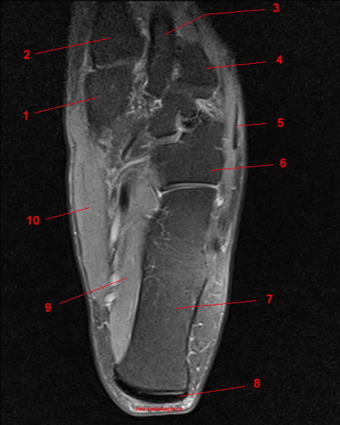
Module 5 Pelvis Imaging from www.hitachihealthcare.com Human muscles enable movement it is important to understand what they do in order to diagnose sports injuries and prescribe rehabilitation exercises. Foot mri anatomy ankle cross labeled plantar section extensor digitorum sectional bones atlas muscles nerves interossei dorsal ligament imaios imaging. The muscular system is an organ system consisting of skeletal, smooth and cardiac muscles. The medial muscles of the foot sole have various tasks: Head, neck, arm, foot, pelvis, etc. Near normal foot mri for reference. 12 photos of the foot muscle anatomy mri. They act collectively to stabilise the arches of the foot, and individually to control movement of the digits.
Structures of the foot shown in this illustration are:
Editor · aug 14, 2017 ·. Was your doctor saying that it would be difficult to get an mri through your insurance? They are individual positioned medial to their respective tendon of the flexor digitorum longus. 12 photos of the foot muscle anatomy mri. Neuropathies around the elbow joint. The calf muscles, including the gastrocnemius and soleus, join to form the strong calcaneal (achilles) tendon. Mri of the ankle and feet. Learn anatomy faster and remember everything you learn. Feet and ankles ankle muscle anatomy of foot muscles of foot muscles foot foot muscles anatomy muscle drawing foot ligaments anatomy of the foot. Related posts of foot muscle anatomy mri muscle anatomy interactive. Near normal foot mri for reference. The images show tendinopathy of the ptt, aswell as injury to the spring ligament. Tendinous, ligamentous, and muscle abnormalities.
They act collectively to stabilise the arches of the foot, and individually to control movement of the digits. Here we explain the major muscles of the human body. Located inferior to the knee are a number of muscles that move the ankle, foot, and toes. Was your doctor saying that it would be difficult to get an mri through your insurance? Involved early gray = muscle:
Mri Of The Ankle Detailed Anatomy W Radiology from w-radiology.com Head, neck, arm, foot, pelvis, etc. Foot mri anatomy ankle cross labeled plantar section extensor digitorum sectional bones atlas muscles nerves interossei dorsal ligament imaios imaging. Common questions and answers about foot anatomy mri. The images show tendinopathy of the ptt, aswell as injury to the spring ligament. The foot contains many bones, muscles, tendons, and other structures. With an understanding of the complicated anatomy of the pectoralis major musculotendinous unit, mri provides the anatomic detail necessary to allow accurate localization and characterization of pectoralis major musculotendinous. The muscles are located mainly in the sole of the foot and divided into a central (medial) group and a group on either side (lateral). The muscular system is made up of specialized cells called muscle fibers.
First of all they act upon the metatarsophalangeal joint of the big toe, leading to the abduction (abductor hallucis muscle), adduction (adductor hallucis muscle) and flexion (both flexor hallucis brevis and adductor hallucis.
The medial muscles of the foot sole have various tasks: The muscles are located mainly in the sole of the foot and divided into a central (medial) group and a group on either side (lateral). Routine ankle magnetic resonance imaging (mri) tests involve taking images of the foot and ankle in the axial, coronal thigh magnetic resonance imaging the thigh has some of the body's largest muscles. Variants, accessory muscles and ossicles. I would guess the referring doctor would have to take that up with them. The calf muscles, including the gastrocnemius and soleus, join to form the strong calcaneal (achilles) tendon. Learn anatomy faster and remember everything you learn. The foot contains many bones, muscles, tendons, and other structures. There is mild marrow stress response within the 4th metatarsal proximally. Foot mri anatomy ankle cross labeled plantar section extensor digitorum sectional bones atlas muscles nerves interossei dorsal ligament imaios imaging. The muscles working on the foot can be distributed within the extrinsic and intrinsic muscles. When the muscles tighten (contract) they pull on the tendons, which in turn move the bones. Pectoralis muscle mri & anatomy.
They act collectively to stabilise the arches of the foot, and individually to control movement of the digits foot muscles mri. Their main function is contractibility.